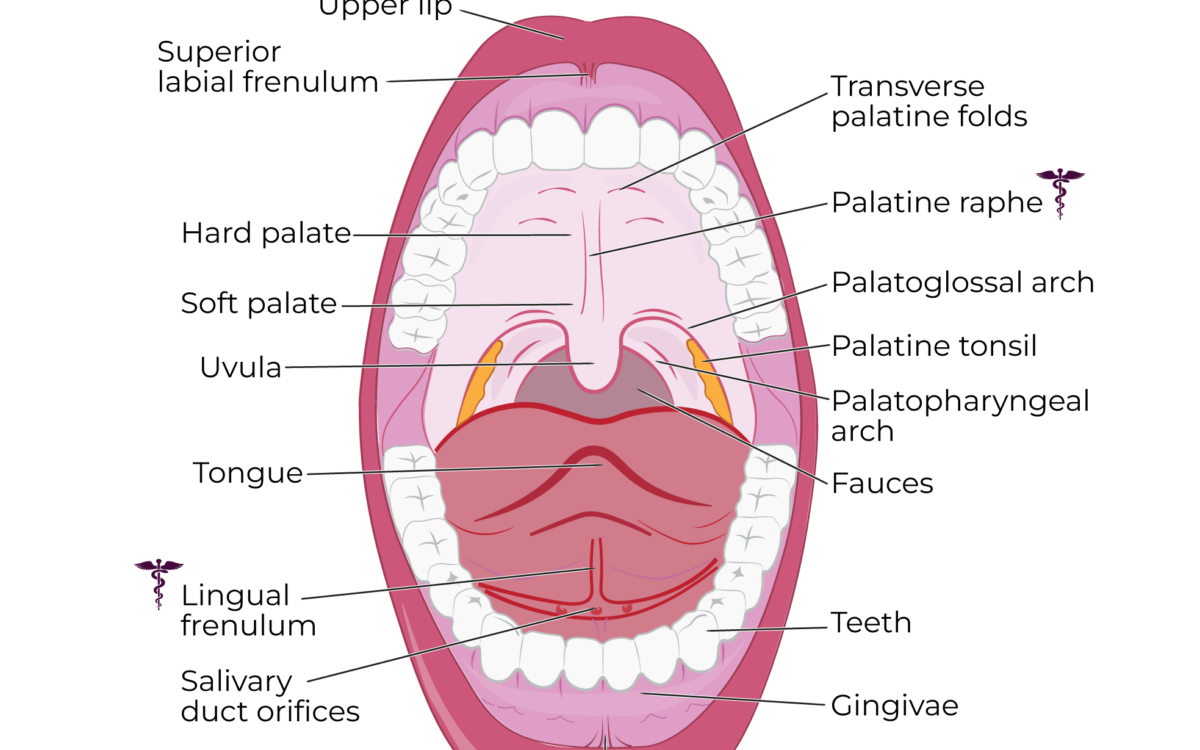
Mouth anatomy is the study of the various structures that make up the oral cavity, which runs from the lips (the oral fissure) at the front to the isthmus of the fauces at the pharynx (throat) at the back. It has two divisions: the oral vestibule, which is the space between lips, cheeks and the gums and teeth, and the oral cavity proper — plus the teeth, gums, palate, tongue, oral mucosa and salivary glands.
This is the short answer. Most answers stop here. This guide continues to provide the real numbers for every structure, their blood supply and nerves, their function, and tells you exactly where the mouth ends and throat begins. It also provides a correction to the tongue taste map that anatomy pages still print.
What is the mouth, and what are its two divisions?
The oral cavity starts at the lips (oral fissure) and ends at the isthmus of the fauces (the pharynx). Between these two structures is the mouth, which is the entrance to the digestive tract. It is also a backup airway. Anatomically it is precise: it begins at the oral fissure — the opening between your lips — and ends at the oropharyngeal isthmus, also called the isthmus of the fauces, the archway at the back where the mouth hands over to the throat.
Inside that span sit two distinct spaces.
The oral vestibule is the space between the cheeks and lips, and the teeth and gums. It is the space a toothbrush sits in. Push your tongue behind your lower lip and you are in it.
The oral cavity proper is bounded by the dental arches and nearly filled by the tongue. TeachMeAnatomy describes the same two-part division, with the cavity running from the oral fissure forward to the oropharyngeal isthmus behind.
Where the vestibule and the cavity proper meet
With your teeth together, these spaces seem to meet, but they don’t. Behind your last molars, at the retromolar trigone, they connect. This is why with your jaw closed you can still sip water and swallow it; the liquid is in the vestibule and passes around the back of the molars and enters the cavity proper.
Mouth anatomy at a glance: the two divisions and six boundaries
This all takes a little time to learn, but with a mirror, a labeled diagram and a few tries, it starts to click.
- Anterior (front): the lips
- Lateral (sides): the cheeks
- Superior (roof): the hard palate in front, the soft palate behind
- Inferior (floor): the floor of the mouth and the tongue
- Posterior (back): the oropharynx
These limits come directly from the clinical reference: StatPearls on the oral cavity, which sets the limits of the cavity as the lips to the isthmus of the fauces, with the hard palate and the soft palate forming the anterior and the posterior roof respectively.
Keep one thing in mind as you read the rest. There are many design elements in your body that are based on function. The mouth has the most of any region in the body due to the abrasive forces from chewing and the temperature extremes from hot and cold food and beverages. The high level of acidity in food and the presence of a resident bacterial population add to the challenges the mouth has to endure. Almost every design feature below, from the keratin on your hard palate to the sheer number of arteries feeding the area, is an answer to these challenges.
What are the parts of the mouth, and what does each part do?
Lips, cheeks and the buccinator
Lips are muscles that frame the oral fissure and seal the mouth. That seal is what makes sucking, swallowing and consonant sounds like /p/ and /b/ possible. Without them, and especially if they are weak on one side, both eating and speech degrade immediately.
The muscle in the cheek, the buccinator, helps push the cheek against the teeth, and its job is underrated. This helps hold food on the surface of the teeth for chewing instead of collecting in the vestibule. When the buccinator weakens, food collects in the cheek — a sign clinicians look for.
Teeth and gums
Teeth have four mechanical functions; incisors cut, canines tear, premolars crush, molars grind. Britannica defines this system as a means to break food down small enough to be digested.
The counts are specific. Colgate puts most adults at 32 permanent teeth — 8 incisors, 4 canines, 8 premolars and 12 molars, the last 4 being wisdom teeth. Children have 20 primary teeth: incisors, canines and molars, but no premolars at all.
Gingiva is the keratinised collar of mucosa that seals around each tooth and covers the alveolar bone underneath. It is firm to the touch, does not bleed during tooth brushing, and ranges from pale pink to brown depending on your skin tone. The gingival margin — the thin rim right where gum meets tooth — is the structure that fails first in gum disease. For this reason, bleeding along the margin is an early signal rather than a minor one.
Hard palate, soft palate and uvula
The roof of your mouth is two different things. The hard palate is the boney section of the roof of the mouth and the soft palate is the muscular section located just behind the hard palate. The soft palate contains five muscles, two of which are the levator veli palatini and tensor veli palatini.
Together they form the dividing wall between the nasal and oral cavities. Britannica makes the functional point well: the palate gives you separate passageways for air and food, which is what lets you breathe and chew at the same time. During a swallow, the soft palate and uvula lift and close off the nasopharynx to prevent any fluid from going down the nasal passage. This is why drink doesn’t go down your nose — and why it sometimes does when you laugh mid-swallow and the timing slips.
The tongue, papillae and the taste map myth
Your tongue is a mass of intrinsic and extrinsic muscle, anchored to the floor of the mouth by the lingual frenulum. There are many types of papillae on your tongue: filiform, fungiform and circumvallate. It does four jobs: it positions food for chewing, propels the bolus when you swallow, carries taste, and acts as the body’s most mobile speech articulator.
Now the correction. The different myths that people believe about your tongue are vastly incorrect. For example, your teachers probably told you that your tongue is divided into areas that sense different tastes — sweet at the tip, salty and sour along the sides, bitter at the back. This is not true. All types of taste receptors are present all over your tongue, and the whole tongue senses them more or less equally. According to Live Science’s debunking of the tongue map, the belief of different areas of the tongue sensing different tastes is due to mistranslation and/or oversimplification of David Hänig’s 1901 German doctoral dissertation. Hänig’s data showed only slight differences in sensitivity around the tongue and never showed different areas that sense different tastes. Virginia Collins disproved the myth in 1974 by demonstrating that all tastes are sensed all over the tongue. The chart is still taught and reprinted anyway.
Floor of the mouth and oral mucosa
The floor of your mouth is a muscular sling formed by the mylohyoid and geniohyoid muscles, and the tongue is supported by it. The submandibular and sublingual gland ducts open on the floor of the mouth, which is why you can watch saliva well up under your tongue in a mirror.
The oral mucosa is the protective soft-tissue lining covering everything in the mouth except the enamel of your teeth. Cleveland Clinic describes it exactly that way. What consumer pages tend to skip is that the lining is not uniform. The gingiva is keratinized, as is the hard palate, where chewing abrades it. The lips, cheeks and floor of the mouth are non-keratinised and more flexible. Different areas of the mouth have different turnover rates, rather than being one number for the whole mouth, and this plays a role when considering how long it takes different areas of the oral mucosa to heal — a detail that matters for the healing question further down.
Salivary glands and what saliva actually does
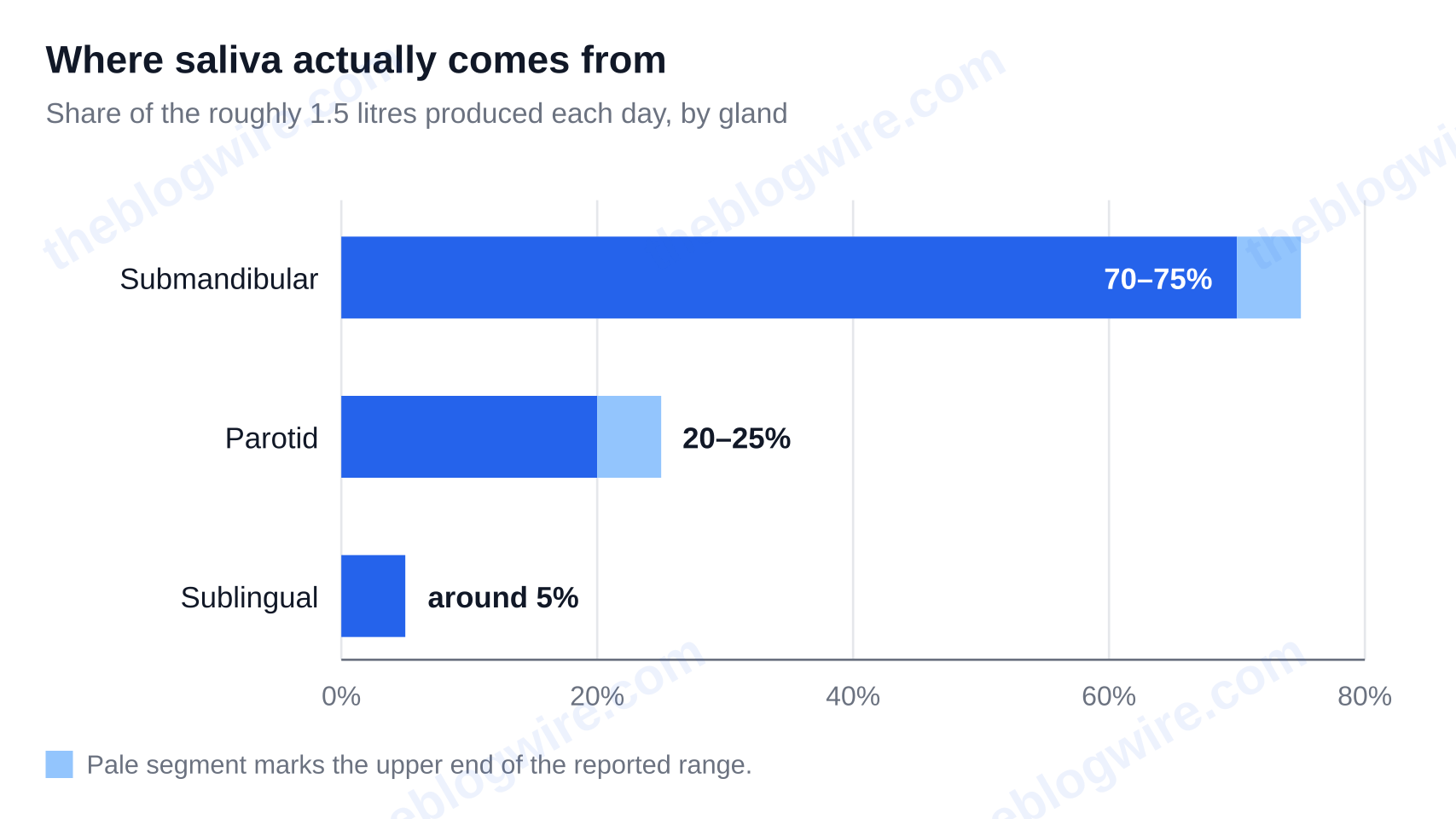
There are 3 paired major salivary glands, and they are not interchangeable. The parotid secretion is purely serous and amylase-rich. The submandibular is mixed serous and mucous. The sublingual is predominantly mucous.
The volumes are lopsided in a way that surprises people. TeachMePhysiology puts total daily volume at roughly 1.5 litres a day. About 70–75% of the total volume is from the submandibular glands, 20–25% from the parotid glands, and around 5% from the sublinguals. The parotid — the one most people can name, and the one mumps swells — contributes a fifth.
Behind the major salivary glands are the minor salivary glands, which most mouth anatomy pages simply wave at with a vague “numerous glands surround the mouth”. The actual numbers, from a review of the major and minor salivary glands: about 800–1,000 minor glands, each only 1–5 mm across, and they have no true capsule. About half of them are located in the mucosa of the hard palate. The minor salivary glands are absent in the gingiva, from the midline and anterior hard palate, and the anterior dorsum of the tongue.
Saliva performs four main functions: starts starch digestion with amylase, lubricates food and aids in speech, washes away food debris and bacteria, and buffers acid. Remove it and all four of these functions fail together — which is the mechanism behind most dry-mouth damage.
Which nerves and blood vessels supply each part of the mouth?
This information is extremely hard to find. The public pages that rank high for this topic name the parts and their functions, however, have no innervation whatsoever, and the clinical pages give the nerve and artery detail in paragraph format. Therefore, to actually obtain the mapping, a person has to gather information from at least 3 sources. So here it is as one lookup table.
| Structure | Sensory supply | Motor supply | Arterial supply | Note |
|---|---|---|---|---|
| Upper lip, labial and buccal mucosa | Maxillary division of trigeminal (V2) | Facial nerve (VII) to the surrounding muscles | Facial artery branches | Sensation and movement split between two different cranial nerves |
| Upper teeth and gingiva | V2, superior alveolar nerves | — | Superior alveolar arteries | Teeth have sensory supply only |
| Hard palate | V2, greater palatine nerve | — | Greater palatine artery | Keratinised, and holds about half the minor salivary glands |
| Lower lip, lower teeth and gingiva | Mandibular division of trigeminal (V3), inferior alveolar nerve | Facial nerve (VII) to the lip musculature | Inferior alveolar artery | One nerve block covers teeth, lip and chin together |
| Floor of the mouth | V3, lingual nerve | V3 to mylohyoid; C1 via hypoglossal to geniohyoid | Lingual artery branches | Submandibular and sublingual ducts open here |
| Cheek and buccinator | V3, buccal nerve | Buccal branches of the facial nerve (VII) | Facial and buccal arteries | Feeling and movement come from different nerves — a classic exam point |
| Anterior two-thirds of tongue | Taste: chorda tympani of the facial nerve (VII). General sensation: lingual nerve of V3 | Hypoglossal nerve (XII) | Lingual artery | Three cranial nerves serve one piece of tongue |
| Posterior third of tongue | Both taste and general sensation: glossopharyngeal nerve (IX) | Hypoglossal nerve (XII) | Lingual artery | Anatomically oropharynx, not oral cavity |
| Tongue muscles | — | Hypoglossal nerve (XII), except palatoglossus | Lingual artery | Palatoglossus is the single exception — supplied by the vagus |
| Soft palate | Lesser palatine branches of V2 | Vagus via the pharyngeal plexus, except tensor veli palatini from V3 | Lesser palatine and ascending palatine arteries | Five muscles; one of them breaks the pattern |
| Salivary glands | — | Secretomotor: parotid via IX; submandibular and sublingual via VII | External carotid branches | Gland control runs on different nerves than the mucosa around them |
How to remember it: four rules instead of twenty facts
The table is extremely lengthy. The information behind it is very short. Four rules cover it almost entirely:
- General sensation for the oral cavity is the trigeminal nerve. The upper half is V2 and the lower half is V3 and there are very few exceptions to this.
- Front-to-back: taste splits 2/3 facial nerve (via chorda tympani) and 1/3 glossopharyngeal nerve.
- Tongue movement is via the hypoglossal cranial nerve — except for palatoglossus, which is via vagus.
- All the arteries come off the external carotid.
The lingual, facial and maxillary arteries are the ones we should know and some of their branches: greater palatine, superior alveolar and inferior alveolar. Blood drains via the facial and lingual veins and the pterygoid venous plexus.
Three practical payoffs fall straight out of the table. An inferior alveolar nerve block numbs the lower teeth, lip and chin together, as one nerve carries all three. Your tongue can feel normal while taste is altered after middle ear surgery, because the chorda tympani runs through the middle ear and sensation does not. And the mouth bleeds freely but has a good, rapid healing response — that dense external carotid supply cuts both ways.
Where does the mouth end and the throat begin?
There is a distinct border and most people place it incorrectly.
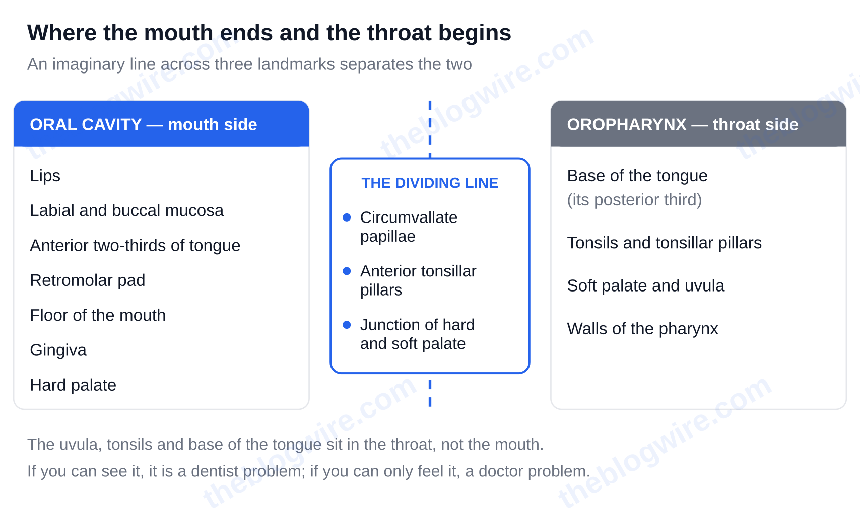
The oral cavity is separated from the oropharynx by an imaginary line running across three landmarks: the circumvallate papillae, the anterior tonsillar pillars, and the junction of the hard and soft palate.
On the mouth side of that line: the lips, labial and buccal mucosa, the anterior two-thirds of the tongue, the retromolar pad, the floor of the mouth, the gingiva and the hard palate. On the throat side: the base of the tongue (its posterior third), the tonsils and tonsillar pillars, the soft palate and uvula, and the walls of the pharynx.
Read that again, because it is counter-intuitive. A careful review of the anatomy indicates the uvula, tonsils and the back of your tongue — structures nearly every consumer anatomy page lists under “parts of the mouth” — are actually in the throat and not in the mouth.
Why the boundary changes the cancer risk profile
This is where the line stops being academic. Tobacco and alcohol are the main drivers of oral cancers, which are squamous cell carcinoma in more than 90% of cases, as noted by StatPearls. HPV-associated cancers are different in that they cluster in the oropharyngeal subsites, which include the tonsils, base of tongue, soft palate and uvula, and the pharyngeal walls — as the CDC’s page on HPV and oropharyngeal cancer describes.
So “mouth cancer” and “HPV throat cancer” are not two names for one disease. They are different diagnoses, with different risk factors, different screening routes and different outlooks, separated by a line you can roughly locate in your own mirror.
Use it that way. A sore in the mouth that won’t heal is on the side of the line that you can see, so it is an oral cavity problem and belongs to a dentist. Persistent one-sided throat or ear pain, a lump in the neck, or difficulty swallowing is on the other side of the line and belongs to a doctor.
What should a healthy mouth look like, and what goes wrong?
What normal looks like inside your mouth
With a mirror and good lighting, your mouth should look something like this. Mucosa should be moist and intact. The gingiva should be firm and not bleeding, with no pockets or recession. The tongue should be pink and have visible papillae. There should be no white or red patches, no ulcer that has outstayed a couple of weeks, no unexplained lump, and enough saliva that your mouth is never sticky when you wake.
There are some things that are alarming and aren’t. A torus palatinus is when there is a hard bony lump in the midline of the hard palate and is a normal variant. The openings of your salivary ducts are supposed to be there, as are the larger papillae across the back of your tongue. People find them while looking for something else and assume the worst.
Common conditions, structure by structure
Grouping conditions by the structures they act on makes them easier to remember than an alphabetical list. Cleveland Clinic’s list of oral conditions lines up neatly with the anatomy:
- Teeth: plaque and cavities
- Gums: gingivitis, periodontitis, recession
- Mucosa: canker sores (aphthous ulcers) and cold sores
- Tongue: glossitis, geographic tongue, tongue tie (a restrictive lingual frenulum)
- Palate: cleft palate
- Salivary glands: dry mouth (xerostomia) and salivary gland tumours
Two mechanisms account for most of that list. First, bacterial biofilm plus a fermentable substrate results in decay and gum disease. Second, anything that reduces saliva, like medication, radiation therapy, dehydration, and an autoimmune disease, removes the buffering and washing system described earlier, and speeds up the first mechanism.
The care that actually changes outcomes is not very glamorous: brush your teeth twice daily with fluoride toothpaste, clean between the teeth, see the dentist regularly, and manage the two main modifiable risk factors for oral cancer, which are tobacco and alcohol.
When to see a dentist or doctor
Stop self-monitoring and book an appointment for any of the following:
- An ulcer, or a white or red patch, lasting more than two to three weeks
- A lump or thickening anywhere in the mouth or neck
- Teeth that have loosened with no injury to explain it
- Persistent bleeding, or numbness that does not resolve
- Difficulty in swallowing, or painful swallowing
- Hoarseness that does not settle
Pull it together and the whole structure is manageable; two divisions, roughly nine structures, each with a job, a nerve and an artery, bounded in front by your lips and behind by a line across the circumvallate papillae and tonsillar pillars. Knowing where that line sits is the difference between a dental Problem and a throat problem. Use the diagram and nerve and artery table above and take yourself through the structures in a mirror, and if anything has been there for longer than two to three weeks, book a review.
As a general rule, if you can see it, it’s a dentist problem. If you can only feel it, it’s a doctor problem. There’s plenty more to dig into on The Blog Wire.
FAQ
Why do cuts and burns inside the mouth heal faster, and with less scarring, than the same injury on skin?
The lining of the mouth replaces itself more quickly. The oral epithelium has a faster cell production rate and a quicker turnover time than the skin. According to wound-healing research published on the NCBI PMC archive, the buccal mucosa replaces itself in 1 to 3 weeks, compared to roughly 4 to 10 weeks for the skin, depending on which part of the skin is being referenced. That faster turnover underpins the oral mucosa’s capacity for rapid, near-scarless healing. It is also important to note that the turnover time varies by site within the mouth too, so the lining on your hard palate and the lining inside your cheek do not behave identically.
Which nerve does a dentist numb for a filling or an extraction?
It depends on which jaw. For lower teeth, the dentist blocks the inferior alveolar nerve, a branch of the mandibular division of the trigeminal nerve (V3). Since this one nerve carries sensation for the lower teeth, the lower lip and the chin, a single injection numbs all three together, which is why your lip feels fat and foreign for an hour afterwards. For the upper teeth, the target is the superior alveolar nerves from the maxillary division (V2), usually infiltrated locally rather than blocked, so the numbness stays more contained.
How many bacteria live in a healthy mouth?
There are far more bacterial species in the mouth than most people imagine. The Human Oral Microbiome Database, described in the Journal of Bacteriology, covers approximately 700 predominant bacterial species of the human oral cavity and includes 619 taxa across 13 phyla. Of those, roughly 280 oral species have been isolated in culture and formally named. The average person carries about 200 species at any given time — a subset of the total, not the whole catalogue. Most of them are normal residents, not invaders; trouble starts when biofilm builds up and meets a steady supply of fermentable sugar.
What causes a bump on the roof of the mouth?
The most common harmless cause of a bump on the roof of the mouth is a torus palatinus — a bony growth in the midline of the hard palate that is a normal anatomical variant, not a disease. Other frequent and benign causes are burns from hot food, canker sores, and blocked minor salivary glands, which makes sense given that around half of the mouth’s 800–1,000 minor salivary glands sit in the mucosa of the hard palate. A good rule of thumb is that if there is a lump, bump or thickening in the mouth that does not go away in two to three weeks, it needs to be checked by a dentist or doctor.