Eversion is the outward rotation of the sole of the foot, with the lateral border of the foot lifting away from the midline. This movement occurs at the subtalar and intertarsal joints and is produced by the fibularis (peroneal) muscles. Eversion is the direct opposite of inversion.
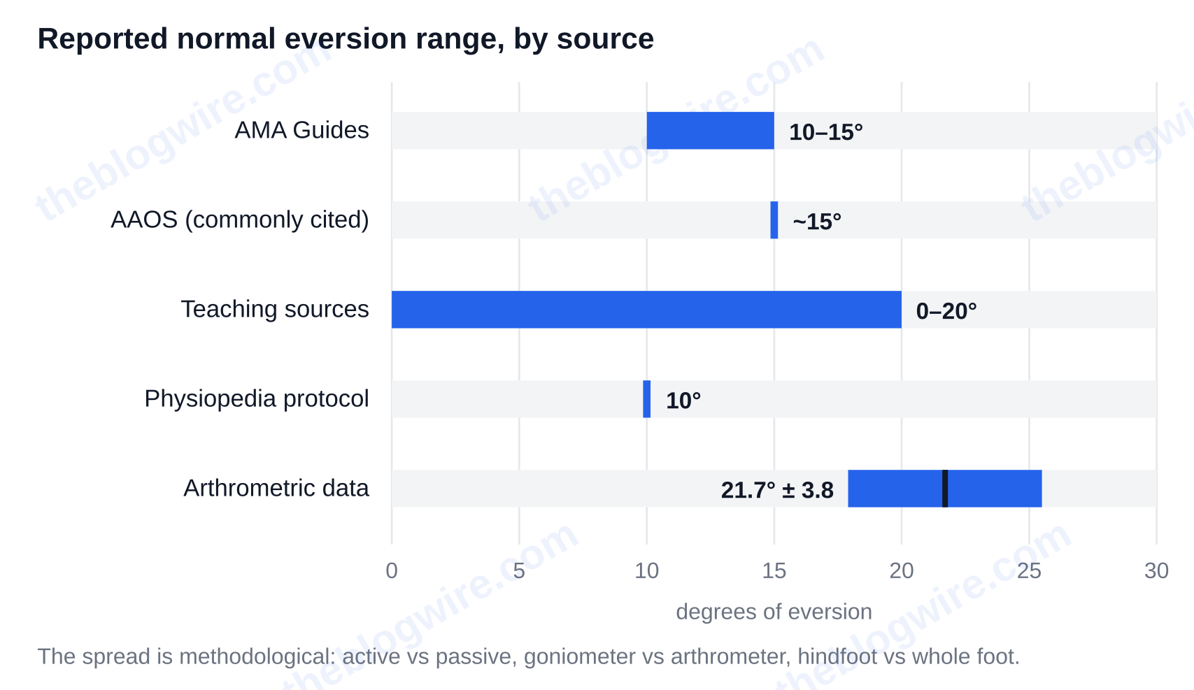
Now the number that makes this topic messy: arthrometric normative data report a mean of 21.7 degrees ± 3.8 for that movement, while the AMA Guides to the Evaluation of Permanent Impairment allow roughly 10–15 degrees. Both describe healthy ankles. The figures differ because the measurement method differs, and that one fact explains most of the contradictions you will hit while revising. Work through the steps below and you will be able to name the joint, the three muscles and the two nerves involved, measure the movement on your own foot in about five minutes, and tell a normal result from one worth getting checked.
Before you start: which meaning do you need?
You will need a chair, a bare foot, and a goniometer or a free phone app to measure angles. You will need five minutes to complete the practical parts of this lesson.
Check that you have the right article, because the bare word has several, and usually divergent, senses. Google’s first result for it is a disambiguation page. Most importantly for your current use, the term has a prominent sense in anatomy and kinesiology. It describes a movement of the foot, and therein lies the sense you need. In differential topology, a similar sense is the turning of a sphere inside out. It is called eversion, and, as Smale’s theorem demonstrates, it is possible. However, in the late 1950s, when the theorem was proven, one could not physically demonstrate the sphere eversion. In ophthalmology, eversion is the turning out of the lid margin, usually of the lower lid, and is called ectropion. In zoology, eversion is the autotomy of a sea cucumber, which ejects its internal organs to defend itself, and the way a starfish turns its stomach outward to digest prey outside its body. Fictionally, it is Alastair Reynolds’s novel, published on 26 May 2022, and the 2010 platform game by Guilherme S. Tows. The root verb ties them all. To evert means to turn outward or inside out.
Step by step: trace eversion from joint to nerve
- With one knee bent, lift the foot clear of the floor. Keeping the leg still, roll the foot so the sole faces away from the other leg. The outer border lifts, and the plantar surface turns laterally. That is eversion. Unlike plantarflexion or dorsiflexion, which are in the sagittal plane, eversion is a frontal (coronal) plane movement like abduction and adduction.
- Write down the joint: the subtalar (talocalcaneal) joint, which is assisted by the intertarsal and tarsometatarsal joints. Cross out “ankle joint” if this is what your notes say. The talocrural joint — the one most people mean by “the ankle” — allows dorsiflexion and plantarflexion.
- Now turn your foot out, once again, and this time press your fingers down the outer side of your lower leg. Under your fingers you will find fibularis (peroneus) longus, from the head and upper lateral fibula, its tendon passing behind the lateral malleolus and under the foot to the first metatarsal and medial cuneiform; and fibularis (peroneus) brevis, from the lower lateral fibula to the base of the fifth metatarsal. Longus assists plantarflexion and aids in the transverse arch. Brevis is the main evertor of the three. Fibularis tertius, which is found in the anterior compartment, assists in the dorsiflexion of the foot and runs to the fifth metatarsal.
- Connect the nerves separately, not as a set. The superficial fibular nerve (L5, S1) supplies longus and brevis, therefore, a lesion to this nerve leaves the outward movement weak or absent. Fibularis tertius is supplied by the deep fibular nerve.
- Contrast the pair out loud: sole turning out, fibularis muscles, smaller range, rarely injured on its own, against sole turning in, tibialis posterior and tibialis anterior, larger range, and cause of the great majority of ankle sprains. Note the mnemonic “outward” describes the sole of the foot, not the leg or knee.
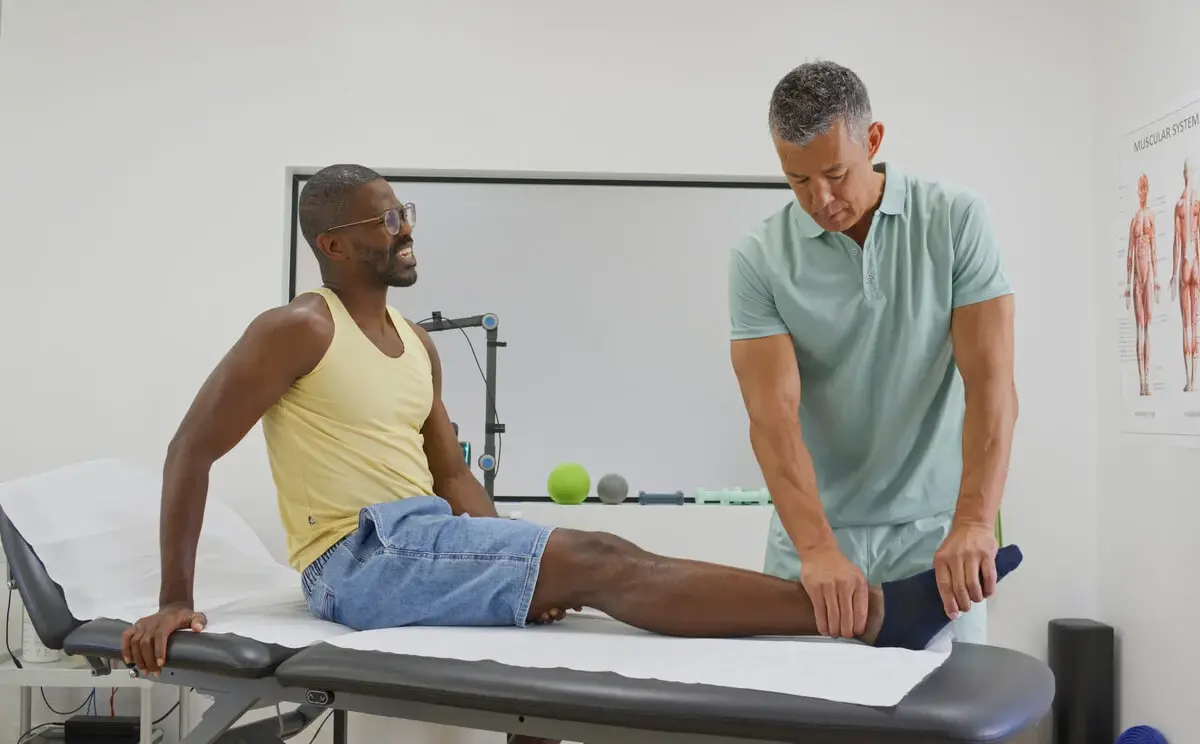
- Measure it. Sit, or lie prone with the knee stabilised. Place the goniometer axis over the back of the ankle, the stationary arm along the midline of the lower leg, and the moving arm along the midline of the calcaneus. Turn the heel outward, and read the angle. Repeat for the other ankle.
Now compare your results to the published figures, which openly disagree:
- AMA Guides to the Evaluation of Permanent Impairment indicates roughly 10–15 degrees of foot eversion.
- AAOS, as commonly cited, indicates about 15 degrees.
- Nursing and anatomy teaching sources indicate 0–20 degrees.
- Physiopedia’s goniometry protocol states an expected range of 10 degrees.
- Arthrometric normative data: a mean of 21.7 degrees ± 3.8, with women measuring higher than men — 16.60 against 13.61 degrees in one dataset.
The spread is methodological, not sloppy: active motion reads differently from passive, a goniometer differently from an arthrometer, and hindfoot-only movement differently from the whole foot. So check your result this way instead of chasing a textbook number. Every source agrees that outward movement gives less range of motion than inversion, and the other ankle is a better reference point. So if the two ankles measure within a few degrees of each other, and they both feel equally resistant to the outward turning force, then you are done.
The one mistake to avoid: calling eversion pronation
These are not synonyms, and saying they are is where marks and clinical reasoning go. Pronation is triplanar. It is an oblique-plane motion at the uniaxial-but-triplanar subtalar joint. It combines the frontal-plane movement above with abduction in the transverse plane and dorsiflexion in the sagittal plane. The outward roll of the sole is only the frontal-plane piece: the principal cardinal-plane component, never the whole motion.
You will find the movement placed “at the ankle joint” or at the intertarsal joints if you look at the results for the bare term. These results flatly contradict one another. The same results list all three fibularis muscles and pass the entire bundle to the superficial fibular nerve, which is incorrect for fibularis tertius — a different compartment and a different nerve. Fibularis tertius may not be present at all; absence is reported from as low as 5% to as high as 72% depending on the population surveyed, which changes what a superficial fibular nerve lesion looks like in front of you. With all this variance, you should trust what you find about the compartment and the nerve more than what you find about the muscle list.
How to strengthen weak evertors — and when the problem is a sprain instead
We can use some variations of banded work to strengthen the evertors. You can anchor a resistance band and loop it around your forefoot, then turn your foot out against the band’s resistance, and return slowly under control. You can also progress to an eccentric loading pattern, which is pulling your foot out to the side and allowing the band to draw the foot back through the range slowly — the loading pattern used in peroneal tendinopathy rehab. You can progress to single-leg balance and perform the work on unstable surfaces, because the reflex that catches an inward roll is what protects you from the common sprain. Weak peroneals leave the ankle unguarded, which is why peroneal strength sits in every sprain rehab plan — and why motion-control shoes and medial-post orthoses exist for feet that roll in too far, since the uncontrolled version of this movement is what “overpronation” and flatfoot describe.
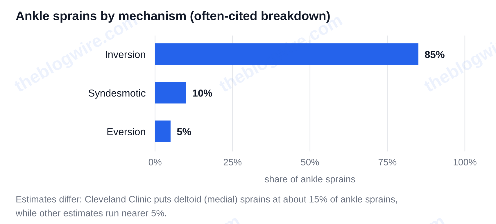
Pain is a different matter. If the foot rolled out and the inside of the ankle hurt and swelled, the suspect isn’t the lateral ligaments. It’s likely the deltoid (medial) ligament complex. These injuries are uncommon, but the published statistics differ widely: Cleveland Clinic puts deltoid or medial ligament sprains at about 15% of ankle sprains, and other estimates run nearer 5%. A breakdown that is often cited is roughly 85% inversion, 5% eversion, and 10% syndesmotic. The low numbers and the lack of consensus is because, in comparison to the lateral ligaments, the deltoid ligament is considerably stronger, and therefore it can take a greater amount of force to tear it. Often, that force will break the medial malleolus or fibula, or injure the syndesmosis instead, and in those cases, combined injuries are classified differently from study to study. Expect medial tenderness below the malleolus and pain on weight-bearing rather than the familiar lateral bruising, expect imaging to rule out fracture first, and expect a lower referral threshold than for a routine inward sprain — a 2024 review in Knee Surgery, Sports Traumatology, Arthroscopy is the current synthesis on deltoid ligament anatomy, diagnosis and treatment. Pain behind the lateral malleolus, an ankle that gives way repeatedly, or outward weakness with no injury behind it all need assessing rather than loading.
Recap: three things to hold onto
First, eversion is a frontal-plane rotation of the sole away from the midline at the subtalar and intertarsal joints. It does not occur at the talocrural ankle joint. Second, eversion is only one plane of pronation and never the whole of it, and the three muscles associated with it are split between two compartments and two nerves. Third, the “normal degrees” figures are in conflict for methodological reasons. Therefore, it is best to compare one ankle to the other rather than relying on a textbook. There’s plenty more to dig into on The Blog Wire.
FAQ
Is eversion the same as pronation?
No. Pronation is a triplanar, oblique-plane motion at the subtalar joint that combines eversion in the frontal plane with abduction in the transverse plane and dorsiflexion in the sagittal plane. Eversion is the principal cardinal-plane component of pronation — one slice of it, not the whole movement.
Does eversion happen at the ankle joint or the subtalar joint?
At the subtalar (talocalcaneal) joint, with the help of the intertarsal and tarsometatarsal joints. The talocrural joint, which most people mean by “the ankle joint”, allows dorsiflexion and plantarflexion. Pages that place eversion “at the ankle joint” are the ones to distrust.
Which nerve controls eversion of the foot?
Fibularis longus and fibularis brevis are supplied by the superficial fibular nerve (L5, S1), so a lesion there leaves eversion weak or absent. An exception is the fibularis tertius muscle: it sits in the anterior compartment and is supplied by the deep fibular nerve.
Can you have an eversion sprain without breaking a bone?
Yes, but it is less likely than with an inversion sprain. The deltoid ligament is considerably stronger than the lateral ligaments, and therefore, an eversion force often fractures the medial malleolus or the fibula, or injures the syndesmosis, instead of producing an isolated sprain. That is why imaging comes first.